


Find out more about how this website uses cookies to enhance your browsing experience.
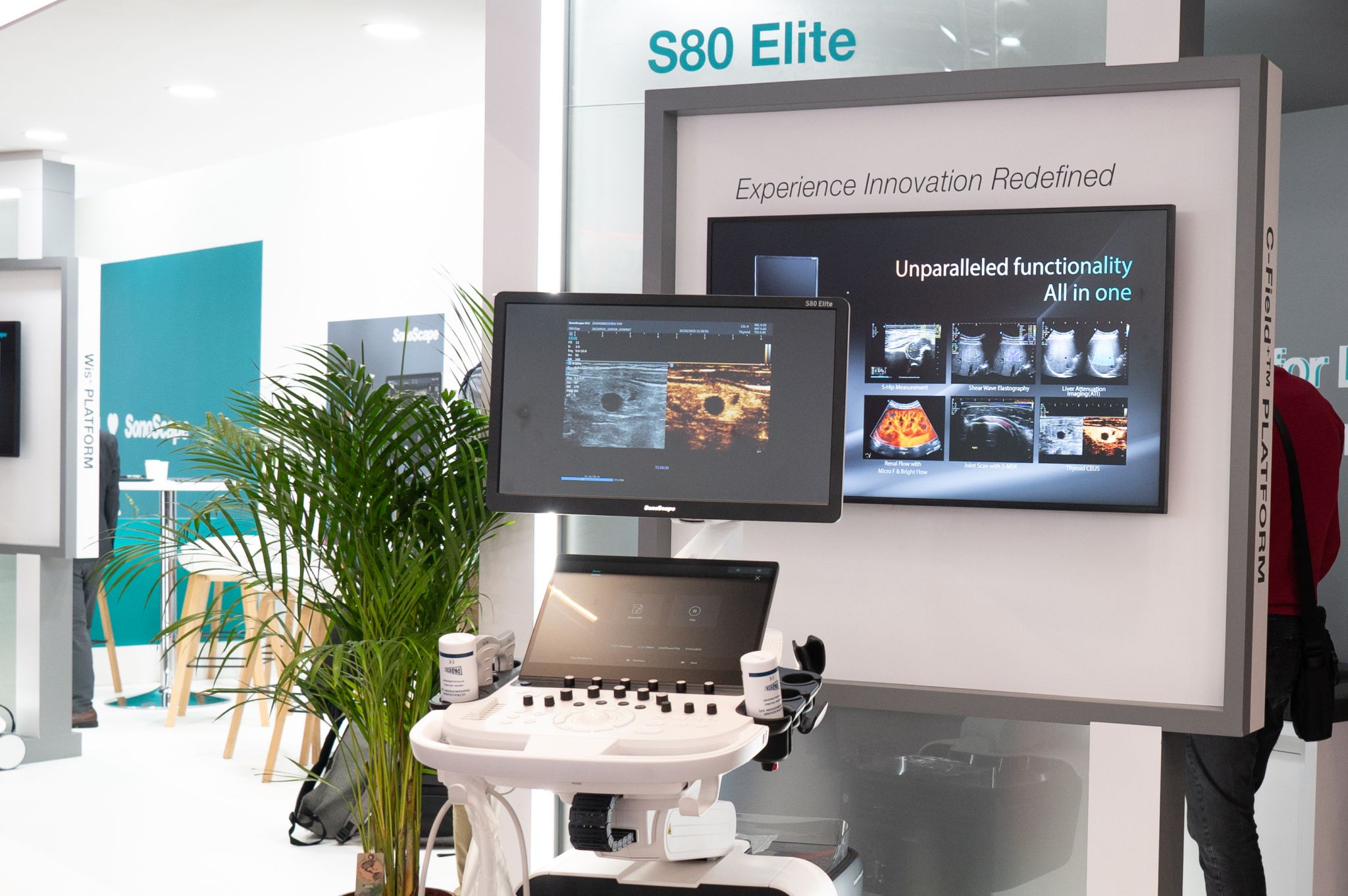
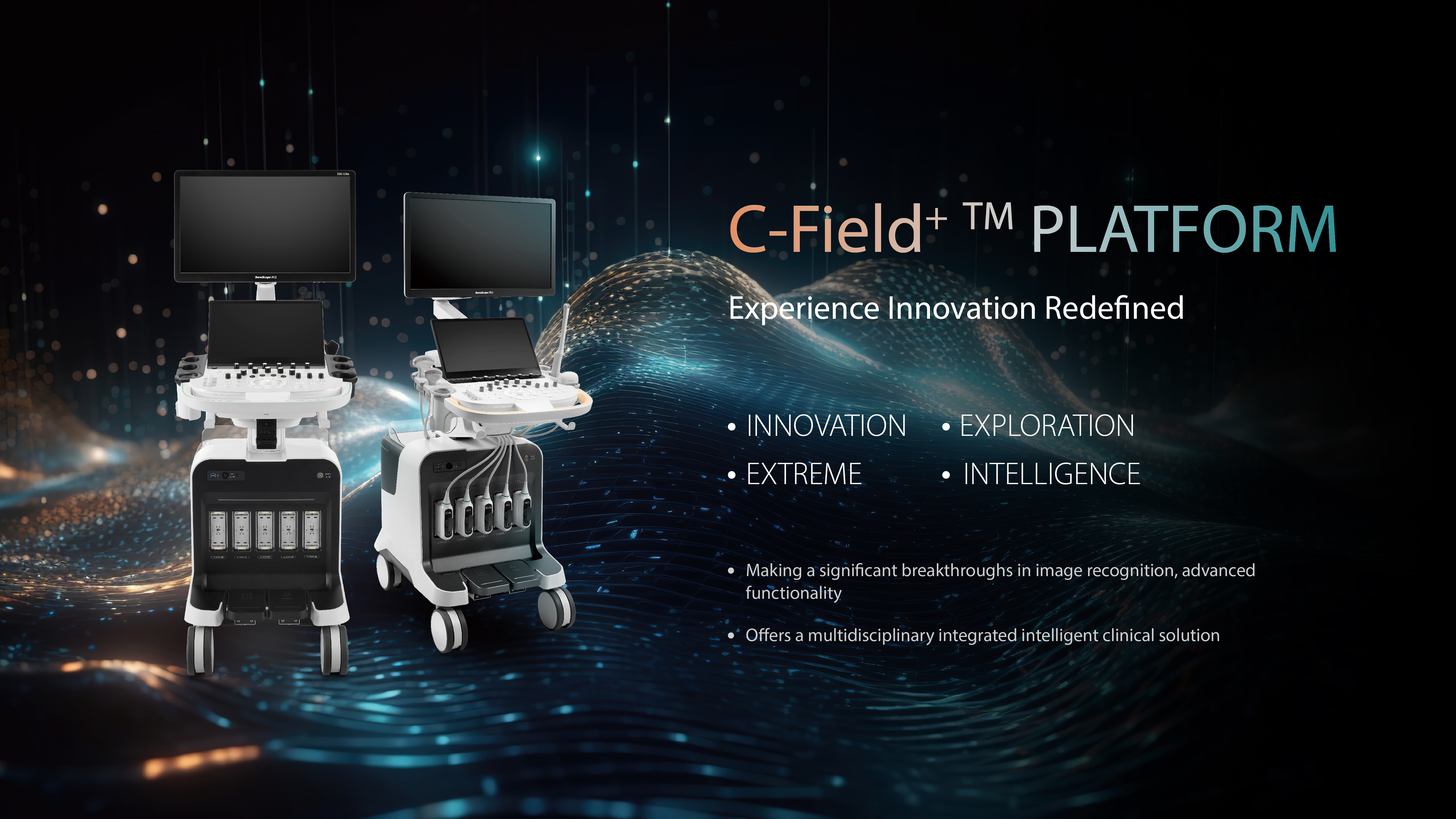
SonoScape S80 Elite
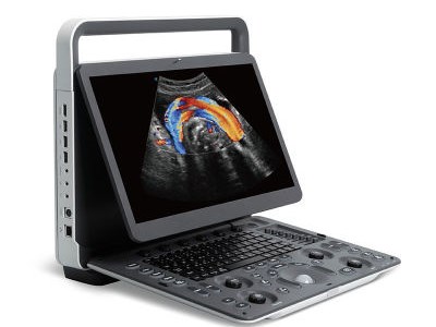
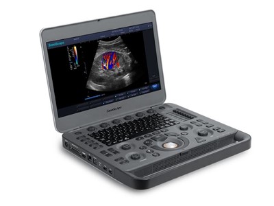
The S80 Elite is a new premium ultrasound product developed by SonoScape based on the C-Field+ platform, making significant breakthroughs in image recognition and advanced functionality. It offers a multidisciplinary integrated intelligent clinical solution. Building upon Sonoscape's AI functionality, the S80 has additional software features to speed up imaging and increase workflow. The system is also capable of advanced imaging functions such as shear wave elastography and fusion imaging.
Available with a range of high definition probes:
- Convex array C1-6A (Abdominal, Obstetrics, Gynecology), 1.0-8.0MHz/ R50mm
- Micro-convex C613 (cardiology, pediatrics), 4-13MHz/ R14mm
- Convex array C322(Abdominal Biopsy), 2-6.8 MHz/ R20mm
- Linear array 12L-A (Vascular, Small parts, MSK etc.), 3-17 MHz/ 52 mm
- Linear array 12L-B (Vascular, Small parts, MSK etc.), 3-17 MHz/ 38 mm
- Phased array 4P-A (Cardiac, Transcranial), 1.0-5.4 MHz
- Phased array 7P-A (Cardiac, Transcranial, Infant),2-9MHz
- Phased array S1-5 (Single crystal) (Cardiac, Transcranial), 1.0-7.0 MHz
- Endocavity 6V3 (Gynecology, Obstetrics, Urology), 3-15 MHz/ R10 mm
- Endocavity 6V3A (Gynecology, Obstetrics), 3-15 MHz/ R8 mm(Vaginal Dilator: EDVS)
- Endocavity 6V7 (Gynecology, Obstetrics), 3-15 MHz/ R10 mm
- Volumetric convex array VC2-9 (Obstetrics, Abdominal, Gynecology), 2-6.8 MHz/ R40 mm
- Volumetric endocavity VE9-5 (Obstetrics, Gynecology, Urology), 2-13 MHz/ R10 mm
- Biplane BCL10-5 (Urology), Convex 3.9-11 MHz/ R10mm, Linear 6-15 MHz/ 60mm
- Phased array transesophageal MPTEE (Cardiac), 4-13 MHz
- Linear array 10I2 (Intra-operative), 4-16 MHz/ 25mm
- Linear array 12LI-A (Intra-operative), 4-16MHz/ 33mm
- Convex array 6CT-A (Intra-operative), 3-15MHz/ R40mm
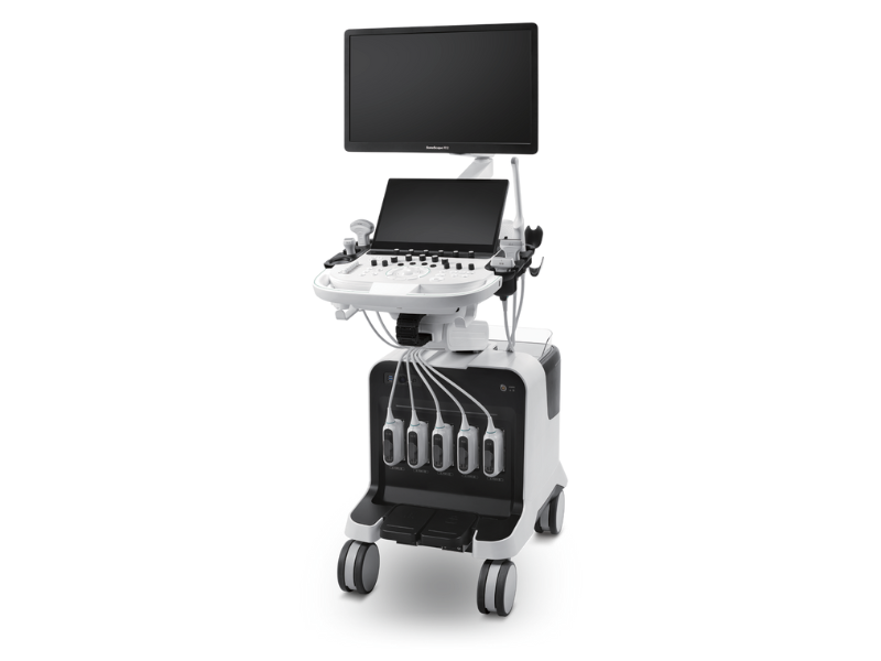
• 24-lnch LED monitor / plus 15-inch tilting touch screen providing a
high definition view and a straightforward workflow.
• Auto' Intelligent assistant package helps to adjust
important imaging parameters (Auto C, Auto PW) and carries out
measurements (Auto EF, Auto Bladder, Auto OB, Auto I MT)
automatically.
• Advanced 3D/4D imaging with increased rendering power for
high clarity visualisation.
• Single Crystal transducers with touch change to enhance
workflow when changing probes.
• Shear Wave Elastography, empowering Quantitative
Assessment for liver fibrosis, thyroid, and breast lesion
characterisation.
• Fatty Liver Attenuation Imaging. The degree of fatty liver
degeneration is quantitatively assessed by calculating the
attenuation coefficient.
• Contrast-Enhanced Ultrasound Imaging. Supports high-frame-
rate contrast imaging to more dynamically display the perfusion
process of lesions.
• Fusion Imaging, with CT/MRI data for simultaneous viewing. Built-in
magnetic sensor in the probe, eliminating the need for an
external magnetic sensor for stand interconnection..
Main Imaging System:
- Digital Beamforming System
- Multiple-beam Parallel Processing Technology
- Digital full focusing technology
- Digital Variable Aperture and Dynamic Aperture Technology
- Digital 2D Grayscale Imaging and M-mode Imaging Unit
- Anatomy M-mode Technology with a minimum of 3 sampling lines, capable of rotating the M-mode sampling line angleby 360 degrees for convenient and accurate measurement.
- Pulse Inversion Harmonic Imaging Unit
- Color Doppler Imaging Technology
- Color Doppler Energy Mapping Technology
- Directional Energy Mapping Technology
- Digital Spectral Doppler Display and Analysis Unit (including PW, CW, and HPRF)
- Intelligent One-Key Image Optimization Technology, capable of adaptively adjusting gain and other parameters to achieve the best image
quality, with a dedicated button.
- Spatial Composite Imaging Technology, supporting multi-level adjustment and combined application of multiple parameters.
- Speckle Noise ReductionTechnology, improving boundary display, enhancing resolution, with at least 5 levels of adjustable grading.
- High-Definition Imaging Technology, sharpening the entire image, enhancing internal tissue resolution and boundary sharpness, with at least 5 levels of independent adjustment.
- Real-time Dual Synchronization/Three Synchronization Function.
- Supports local zoom and one-key full-screen zoom.
- Built-in DICOM 3.0 standard output interface.
- Supports compatibility with workstations of the same brand (proof of registration certificate required).
- WIFI wireless data transmission function, through mobile terminal application software (APP), scan the QR code in the ultrasound device to synchronise and share real-time scanning images on the mobile terminal; can also send image data from the ultrasound device to the mobile terminal for browsing, storage, and smart interaction.
- Workflow protocol, supporting customisable workflow protocol settings, automatically adding annotations, body markers, and activating measurements based on preset processes, combined with a teaching system to help operators smoothly complete examinations.
- Supporting quick report transmission function, allowing reports to be transferred from the ultrasound device to the PC end.
- Supports automatic probe wake-up, with 3 adjustable sensitivity levels.
- Supports voice annotations.
- Supports voice control.
- Supports facial recognition.
- Supports quick switching of diagnostic items.
- Requires the purchased model to be the latest model launched in 2022 or later, with the capability for continuous upgrades.
Contrast Imaging Technology Support
1) Can be used in conjunction with speckle noise reduction technology.
2) Has real-time dual-frame contrast imaging mode, where contrast parameters and 2D parameters can be independently adjusted.
3) Up to 10 minutes of continuous contrast acquisition time.
4) Position interchangeability between contrast images and tissue images.
5) Real-time microvascular contrast imaging technology, which clearly displays the perfusion and trajectory of micro vessels within tissues.
6) Perfusion time imaging technology, based on microvascular contrast imaging, focuses on the time it takes for the contrast agent to reach the vascular cavity. Different colors are used to encode the arrival time of the contrast agent microbubbles in the vascular cavity. Both dynamic videos and static images display the temporal sequence, vascular distribution, and perfusion characteristics of tissue blood flow through different colors within the vessels.
7) Mixed imaging mode of contrast and tissue, displaying a combination of contrast images and tissue images to aid in locating the regions of interest within the tissue.
8) Quantitative analysis of contrast time-intensity curve in contrast imaging mode, supporting calculations and display of 10 Time-Intensity Curves (TIC), automatically calculating perfusion parameters such as arrival time (AT), time to peak (TTP), and peak intensity (PI).
9) In contrast imaging mode, the same probe supports midline, single line, and double line regional puncture guidance functions.
10) Contrast flow imaging technique, which overlays angiographic images with blood flow images, assists doctors in determining the nature of a lesion by combining the angiographic appearance with the blood flow conditions at that location.
Panoramic Imaging Technology
1) Scanning length ≥80cm.
2) Supports measurements.
3) Supports one-click full-screen zoom function.
4) Linear-array probes, convex array probes, and phased array probes all support Panoramic Imaging.
5) Supports color Doppler and energy Doppler (CFM and PDI) real-time Panoramic Imaging.
6) Real-time display of probe movement speed indicator box and speed prompt on the screen at the image stitching area.
Elastography Technology
Support for Strain Elastography
1) Equipped with displacement curves for real-time display of compression frequency and relative displacement magnitude.
2) Built-in integrated real-time elastography quantitative analysis software for area comparison and elasticity comparison analysis of elastography
images.
3) In elastography mode, adjustable colour maps, transparency, contrast, frame correlation, and image optimisation for optimisation of elastography images.
Support for Shear Wave Elastography
1) Supports one-dimensional point shear wave elastography and real-time two-dimensional shear wave elastography.
2) Supports switching between shear wave velocity, Young's modulus, and shear modulus units.
3) Supports quantitative analysis, quantitative analysis ratio, and quantitative analysis histogram.
Support for Fusion Navigation Imaging Technology
Cardiovascular Examination Technology
1) Load echocardiographic analysis, supporting "bull's-eye" plot analysis.
2) Quantitative analysis of myocardial motion, supporting strain, strain rate, velocity, displacement, and volume curve analysis, both local and global myocardial motion quantitative analysis, supporting "bull's-eye" plot analysis.
3) Tissue Doppler imaging and analysis technology (TDI), with various modes including color, PW, and M-mode.
4) Supports automatic measurement of left ventricular and right ventricular functions, as well as automatic strain evaluation.
Thyroid Intelligent Scanning
Technology, with one-button automatic identification of thyroid nodules, automatic marking, measurement, and ultrasound diagnostic analysis of lesions, reducing subjective dependence, good reproducibility, improving work efficiency, and providing guidance, learning, and teaching functions to operators.
Breast Intelligent Scanning
Provides real-time detection and prompt for breast nodules.
With a single button press, it automatically identifies breast nodules and performs automatic marking, measurement, ultrasound diagnostic description, and other analyses on the lesions. This reduces subjective dependence, improves reproducibility, and enhances work efficiency. Additionally, it provides guidance, learning, and teaching functions or operators (Only for tender).
Support for Obstetric Automatic Measurements
1) Supports automatic acquisition of sections.
2) Supports automatic measurements of 12 items in the background, with one-button automatic measurement available for single and four-chamber amniotic fluid sections.
Musculoskeletal Intelligent Scanning Technology
1) In musculoskeletal 2D imaging real-time mode, supports one-button automatic identification of standard bone sections and annotates different tissue structures with different color markings and names.
2) Assists doctors in quickly obtaining standard shoulder joint sections.
3) Supports simultaneous display with the on-machine musculoskeletal teaching system, allowing real-time communication with the machine to help doctors master the techniques for obtaining shoulder joint sections.
Built-in ultrasound teaching software
Providing anatomical diagrams, standard ultrasound images, scanning technique diagrams, and real-time examiner images, instructing operators to perform proper scanning of standard sections, including liver, cardiac, breast, thyroid, kidneys, spleen, uterus, vascular system, biliary system, prostate, gynecology, musculoskeletal and other sections.
Expanded Imaging Technology
Supports convex/convex-micro/linear array probes, with a maximum expansion angle ≥30°, adjustable up to ≥2 level
Accessories:
- B/W Thermal Video Printer: Sony UP-X898MD
- Color Laser Jet Printer: HP M254nw
- B/W Thermal Printer Paper: 1 roll
- Color Thermal Printer Paper: 1 box (80 pages)
- Vaginal dilator
- Blue tooth remote control
- ECG lead wire
- External DVD Drive (USB interface)
- Footswitch
- Gel
Mainly used for clinical diagnosis and teaching in fields such as abdomen, cardiac urology, neonatology, pediatrics, vascular (peripheral, cerebral, abdominal), small organs, musculoskeletal, nerves, contrast imaging, and interventions. The S80 Elite has the capability for continuous upgrades to meet new clinical application requirements.


Training and support
All purchases come with in depth technical training, and ongoing support to ensure you get the best results from your SonoScape scanner. Required pre-sets will be programmed at install.
Servicing
All our scanners require regular servicing, our dedicated team of engineers are able to service units at a time convenient for you to ensure your machine remains in top condition.
Free Demonstration
Celtic SMR is offering free demonstrations of the SonoScape products so please call us on 0800 279 9050 to find out more.
Related Products